Figure 1
Normal Value of Skull Base Angle Using the Modified Magnetic Resonance Imaging Technique in Thai Population
Siriporn Hirunpat*, Nat Wimolsiri and Nuttha Sanghan
Published: 20 March, 2017 | Volume 2 - Issue 1 | Pages: 017-021

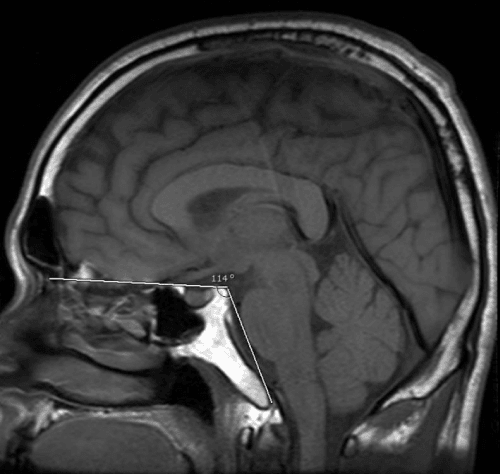
Figure 1:
The angle formed by the line extending across the anterior cranial fossa to the tip of the dorsum sellae and another line drawn along the posterior margin of the clivus in mid sagittal SE T1 weighted image.
Read Full Article HTML DOI: 10.29328/journal.johcs.1001006 Cite this Article Read Full Article PDF
More Images
Similar Articles
-
Infection Control Mechanisms Employed by Dental Laboratories to Prevent Infection of their Dental Technicians/TechnologistsKilimo C Sammy*,Simiyu N Benjamin. Infection Control Mechanisms Employed by Dental Laboratories to Prevent Infection of their Dental Technicians/Technologists. . 2016 doi: 10.29328/journal.johcs.1001001; 1: 001-011
-
Evaluation of ImageJ for Relative Bone Density Measurement and Clinical ApplicationManuel Geiger*,Galina Blem,Arwed Ludwig. Evaluation of ImageJ for Relative Bone Density Measurement and Clinical Application . . 2016 doi: 10.29328/journal.johcs.1001002; 1: 012-021
-
Promising Future in the Detection of Oral Cancer by Using Advance Screening TechnologyMohamed Yasser Kharma*,Mohamed Sadek Alalwani,Manal Fouad Amer. Promising Future in the Detection of Oral Cancer by Using Advance Screening Technology . . 2016 doi: 10.29328/journal.johcs.1001003; 1: 022-33
-
Evaluation of Horizontal Lip Position in Adults with Different Skeletal Patterns: A Cephalometric StudyRohit Kulshrestha*,Vinay V Umale,Kamlesh Singh,Aftab Azam,Madhvi Bhardwaj. Evaluation of Horizontal Lip Position in Adults with Different Skeletal Patterns: A Cephalometric Study . . 2017 doi: 10.29328/journal.johcs.1001005; 2: 009-016
-
Normal Value of Skull Base Angle Using the Modified Magnetic Resonance Imaging Technique in Thai PopulationSiriporn Hirunpat*,Nat Wimolsiri,Nuttha Sanghan. Normal Value of Skull Base Angle Using the Modified Magnetic Resonance Imaging Technique in Thai Population . . 2017 doi: 10.29328/journal.johcs.1001006; 2: 017-021
-
Comparative Study of Enophthalmos Treatment with Titanium Mesh Combined with Absorbable Implant vs. Costochondral Graft for Large Orbital Defects in Floor FracturesMalagón Hidalgo*,Héctor Omar,González Magaña,Fernando, Kalach Mussali,Alberto Jaime,Mejía Valero,Sergio Abraham,Vilchis López,Roberto,Araiza Gómez,Edgardo,Kalach Mussali. Comparative Study of Enophthalmos Treatment with Titanium Mesh Combined with Absorbable Implant vs. Costochondral Graft for Large Orbital Defects in Floor Fractures . . 2017 doi: 10.29328/journal.johcs.1001007; 2: 022-29
-
Visualization and Evaluation of Changes after Rapid Maxillary ExpansionIlija Christo Ivanov*,Dagmar Strakova,Tatjana Dostalova,Jan Dupej,Sarka Bejdova,Veronika Ciganova. Visualization and Evaluation of Changes after Rapid Maxillary Expansion . . 2017 doi: 10.29328/journal.johcs.1001008; 2: 030-37
-
Orthodontics Miniscrews to Correct an Anchorage Loss: Case ReportHoub-Dine A*,Zaoui F. Orthodontics Miniscrews to Correct an Anchorage Loss: Case Report. . 2017 doi: 10.29328/journal.johcs.1001009; 2: 038-042
-
External Root Resorption associated with Impacted Third Molars: A Case ReportRenato Marano*,Gabriela Mayrink,Paula Ramos Ballista,Laisa Kinderlly,Stella Araujo. External Root Resorption associated with Impacted Third Molars: A Case Report . . 2017 doi: 10.29328/journal.johcs.1001010; 2: 043-048
-
Clown language training in Dental education: Dental Student’s PerspectiveSiddharth Tevatia*,Richa Dua,Vinita Dahiya,Nikhil Sharma,Rahul Chopra,Vidya Dodwad. Clown language training in Dental education: Dental Student’s Perspective . . 2017 doi: 10.29328/journal.johcs.1001011; 2: 049-056
Recently Viewed
-
Environmental Factors Affecting the Concentration of DNA in Blood and Saliva Stains: A ReviewDivya Khorwal*, GK Mathur, Umema Ahmed, SS Daga. Environmental Factors Affecting the Concentration of DNA in Blood and Saliva Stains: A Review. J Forensic Sci Res. 2024: doi: 10.29328/journal.jfsr.1001057; 8: 009-015
-
Biotechnology in Forensic Science: Advancements and ApplicationsSunny Antil,Vandana Joon*. Biotechnology in Forensic Science: Advancements and Applications. J Forensic Sci Res. 2025: doi: 10.29328/journal.jfsr.1001073; 9: 007-014
-
Maximizing the Potential of Ketogenic Dieting as a Potent, Safe, Easy-to-Apply and Cost-Effective Anti-Cancer TherapySimeon Ikechukwu Egba*,Daniel Chigbo. Maximizing the Potential of Ketogenic Dieting as a Potent, Safe, Easy-to-Apply and Cost-Effective Anti-Cancer Therapy. Arch Cancer Sci Ther. 2025: doi: 10.29328/journal.acst.1001047; 9: 001-005
-
Buffer Solutions of known Ionic StrengthVíctor Cerdà*, Piyawan Phansi. Buffer Solutions of known Ionic Strength. Ann Adv Chem. 2023: doi: 10.29328/journal.aac.1001043; 7: 051-056
-
Fostering Pathways and Creativity Responsible for Advancing Health Research Skills and Knowledge for Healthcare Professionals to Heighten Evidence-Based Healthcare Practices in Resource-Constrained Healthcare SettingsJanvier Nzayikorera*. Fostering Pathways and Creativity Responsible for Advancing Health Research Skills and Knowledge for Healthcare Professionals to Heighten Evidence-Based Healthcare Practices in Resource-Constrained Healthcare Settings. J Community Med Health Solut. 2025: doi: 10.29328/journal.jcmhs.1001052; 6: 005-019
Most Viewed
-
Evaluation of Biostimulants Based on Recovered Protein Hydrolysates from Animal By-products as Plant Growth EnhancersH Pérez-Aguilar*, M Lacruz-Asaro, F Arán-Ais. Evaluation of Biostimulants Based on Recovered Protein Hydrolysates from Animal By-products as Plant Growth Enhancers. J Plant Sci Phytopathol. 2023 doi: 10.29328/journal.jpsp.1001104; 7: 042-047
-
Sinonasal Myxoma Extending into the Orbit in a 4-Year Old: A Case PresentationJulian A Purrinos*, Ramzi Younis. Sinonasal Myxoma Extending into the Orbit in a 4-Year Old: A Case Presentation. Arch Case Rep. 2024 doi: 10.29328/journal.acr.1001099; 8: 075-077
-
Feasibility study of magnetic sensing for detecting single-neuron action potentialsDenis Tonini,Kai Wu,Renata Saha,Jian-Ping Wang*. Feasibility study of magnetic sensing for detecting single-neuron action potentials. Ann Biomed Sci Eng. 2022 doi: 10.29328/journal.abse.1001018; 6: 019-029
-
Pediatric Dysgerminoma: Unveiling a Rare Ovarian TumorFaten Limaiem*, Khalil Saffar, Ahmed Halouani. Pediatric Dysgerminoma: Unveiling a Rare Ovarian Tumor. Arch Case Rep. 2024 doi: 10.29328/journal.acr.1001087; 8: 010-013
-
Physical activity can change the physiological and psychological circumstances during COVID-19 pandemic: A narrative reviewKhashayar Maroufi*. Physical activity can change the physiological and psychological circumstances during COVID-19 pandemic: A narrative review. J Sports Med Ther. 2021 doi: 10.29328/journal.jsmt.1001051; 6: 001-007

HSPI: We're glad you're here. Please click "create a new Query" if you are a new visitor to our website and need further information from us.
If you are already a member of our network and need to keep track of any developments regarding a question you have already submitted, click "take me to my Query."


